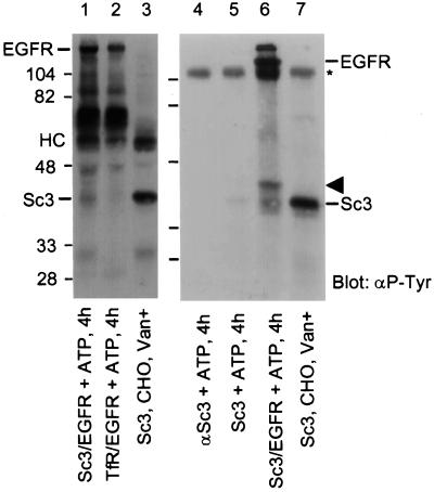
Figure 8.
In vitro phosphorylation of SCAMP3 by EGFR. EGFR was immunoprecipitated from NeoR cells stimulated with 100 ng/ml EGF for 5 min. SCAMP3 and transferrin receptor were immunoprecipitated from CHO cells. Lanes 1 and 6, EGFR and SCAMP3 incubated with cold ATP for 4 h; lane 2, EGFR and transferrin receptor incubated with cold ATP for 4 h. Lanes 3 and 7, SCAMP3 immunoprecipitated from vanadate-treated CHO cells; lane 4, anti-SCAMP3 alone incubated with ATP for 4 h; lane 5, SCAMP3 incubated with ATP for 4 h. HC indicates the position of the IgG heavy chain; * indicates the position of nonreduced IgG. The arrowhead indicates the position of an unknown phosphotyrosine-containing protein. Note that the samples in lanes 1–3 were reduced with DTT before electrophoresis, whereas those in lanes 4–7 were not. Many of the nonspecific bands observed in the vicinity of the IgG heavy chain (HC, lanes 1–3) redistribute to higher apparent Mr (lanes 4–7). All samples were Western blotted for phosphotyrosine and visualized using ECL.

