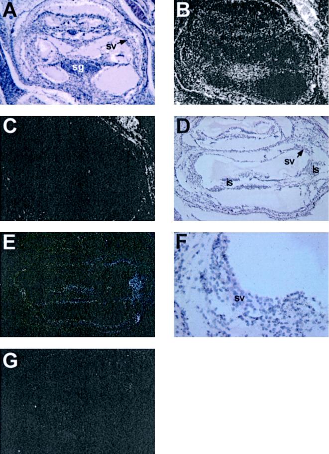
Figure 1.
In situ hybridization analysis of Mpv17 and MMP-2 expression in murine cochleae at 4 d of age. (A–C) Mpv17 expression in wild types: (A) bright field picture; (B) dark field picture of a Mpv17 antisense probe hybridization; (C) Mpv17 sense probe hybridization. Exposure time (A–C), 16 d; magnification 50×. (D–G) MMP-2 expression: (D) bright field picture; (E) dark field picture of a MMP-2 antisense probe hybridization. Exposure time (D-G), 12 d; magnification 50×. (F) Bright field picture of the stria vascularis of panel E. Magnification, 200x. (G) MMP-2 sense probe hybridization. Magnification 50×. sv, stria vascularis; ls, ligamentum spirale; is, inner sulcus; sg, spiral ganglion. The pictures were scanned with a Scan Maker Designer Pro and assembled using Adobe Photoshop 4.0.

