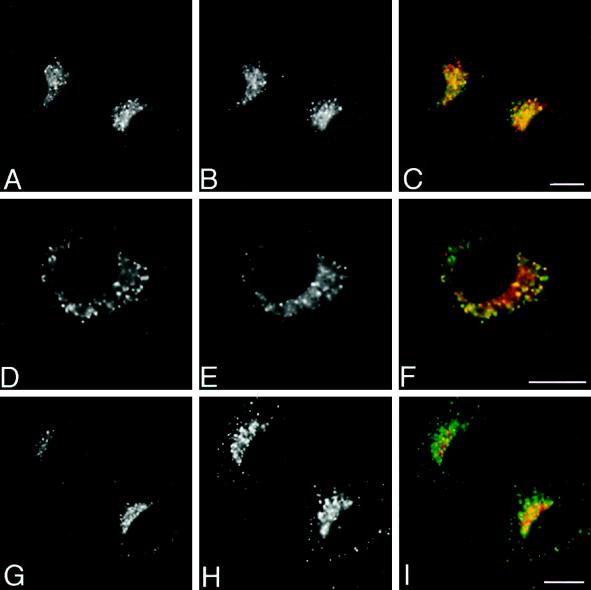
Figure 3.
Immunofluorescence localization of P-selectin to endosomes in NRK cells. NRK cells expressing native P-selectin were processed for immunofluorescence microscopy and double labeled with antibodies to P-selectin (A, D, and G) and either transferrin receptor (B), cation-independent mannose 6-phosphate receptor (E), or lgpA (H) followed by FITC–goat anti-mouse IgG and Texas Red–goat anti-rabbit IgG. The combined images for each double label are shown in C, F, and I. P-selectin was detected in a significant fraction of transferrin receptor-positive structures (A–C) and in some mannose 6-phosphate receptor-positive structures (D–F) and was not detected in lgpA-positive structures (G–I). Bars, 8 μm.

