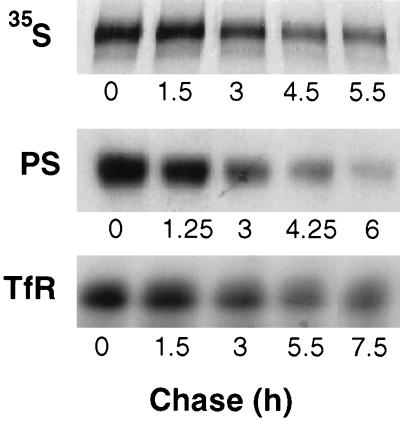
Figure 4.
Turnover of P-selectin and transferrin receptor in NRK cells. Top, metabolic labeling of P-selectin. NRK cells expressing native P-selectin were labeled with [35S]amino acids for 1 h and then chased for 1 h to allow for transport through the Golgi apparatus (t = 0). Plates were then harvested at the indicated intervals. P-selectin was immunoprecipitated from detergent lysates of the cells and separated by SDS-PAGE. Radioactivity was quantitated by PhosphorImager analysis. A radiograph of the gel is shown. Middle and bottom, cell surface labeling. NRK cells expressing native P-selectin were labeled with biotin at 0°C and then recultured for the indicated times before detergent lysis and immunoprecipitation of P-selectin (PS, middle) or endogenous transferrin receptor (TfR, bottom). Precipitates were separated by SDS-PAGE, transferred to Immobilon membranes, and probed with 125I-streptavidin. Autoradiographs of the blots are shown. Radioactivity was quantitated by PhosphorImager analysis. Half-lives were calculated as described in MATERIALS AND METHODS. In these experiments, the half-life of P-selectin was 2.5 h by metabolic labeling and 3.0 h by cell surface labeling, and the half-life of transferrin receptor was 9.0 h.

