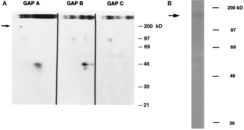
Figure 4.
Complex 1 contains a ∼160-kDa RNA-binding protein. (A) 32P-radiolabeled GAP A, B, and C were incubated with brain protein extract and analyzed on a 6% nondenaturing polyacrylamide gel. The gel was exposed to UV light, and the protein–RNA complexes were visualized by autoradiography. The entire lane in which the reaction was run was excised and layered (as shown in the upper portion of the figure) onto a 10% SDS polyacrylamide gel. The arrow indicates a ∼160-kDa protein associated with complex 1. (B) Binding reaction of GAP A probe and protein extract was electrophoresed through nondenaturing gels and visualized as in panel A. Complex 1 was excised, and the proteins were electroeluted and analyzed on a 10% SDS polyacrylamide gel. The arrow indicates a ∼160-kDa protein associated with complex 1.

