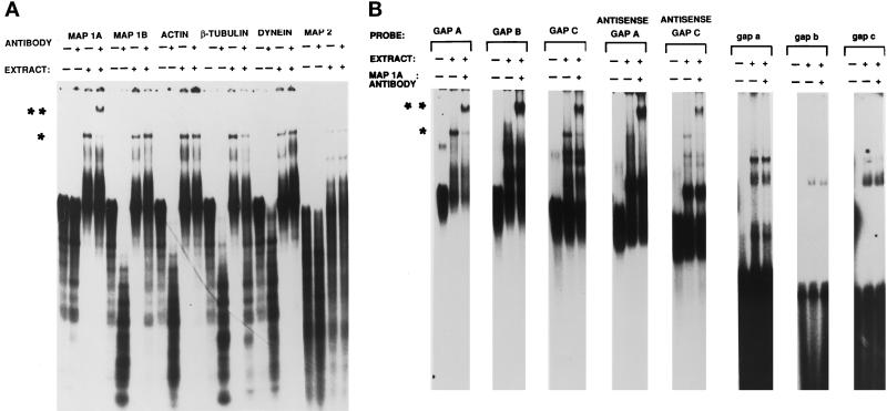
Figure 6.
The microtubule-associated protein MAP 1 A is a component of complex 1. (A) Radiolabeled GAP A was incubated with brain protein extract in the presence of antibodies to several cytoskeletal components and electrophoresed through a 6% nondenaturing polyacrylamide gel. For each probe, control lanes with probe alone, probe with antibody alone, and probe with extract alone are shown to the left of the experimental lanes containing probe, extract, and antibody. The single asterisk indicates the position of complex 1, and the double asterisk indicates the position of the supershifted complex 1. (B) GAP A, GAP B, GAP C, antisense GAP A, antisense GAP C, gap a, gap b, and gap c RNA probes were incubated with brain protein extract in the presence of antibodies that recognize MAP 1A. The first two lanes for each of the gap a, gap b, and gap c probes are identical to those shown in Figure 3c. The single asterisk denotes complex 1, while the double asterisk denotes the supershifted complex 1, indicating the presence of MAP 1A.

