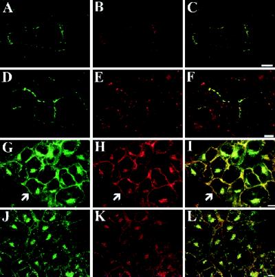
Figure 4.
Colocalization of Cx43-GFP fluorescence with immunofluorescently labeled GFP and Cx43 in NRK and N2A cells. NRK cells (A–F) transiently expressing Cx43-GFP (A and D, green) were immunolabeled with anti-GFP (B, red) or anti-Cx43 (E, red) antibodies. In both instances, all of the GFP fluorescence colocalized with the anti-GFP (C, yellow) or anti-Cx43 (F, yellow) antibody-labeled structures. The additional immunostaining observed when NRK cells were labeled with anti-Cx43 antibodies was mostly likely due to the presence of wild-type Cx43 (E and F, red). The fluorescence of Cx43-GFP in N2A cells stably expressing the fusion protein (G and J, green) completely colocalized with the anti-GFP (H, red) and anti-Cx43 (K, red) immunofluorescent labeling patterns (see yellow in overlays; I and L). The arrows denote cell surface localization of Cx43-GFP in N2A cells where the cell does not have an apposed neighbor (G–I). Bar, 10 μm.

