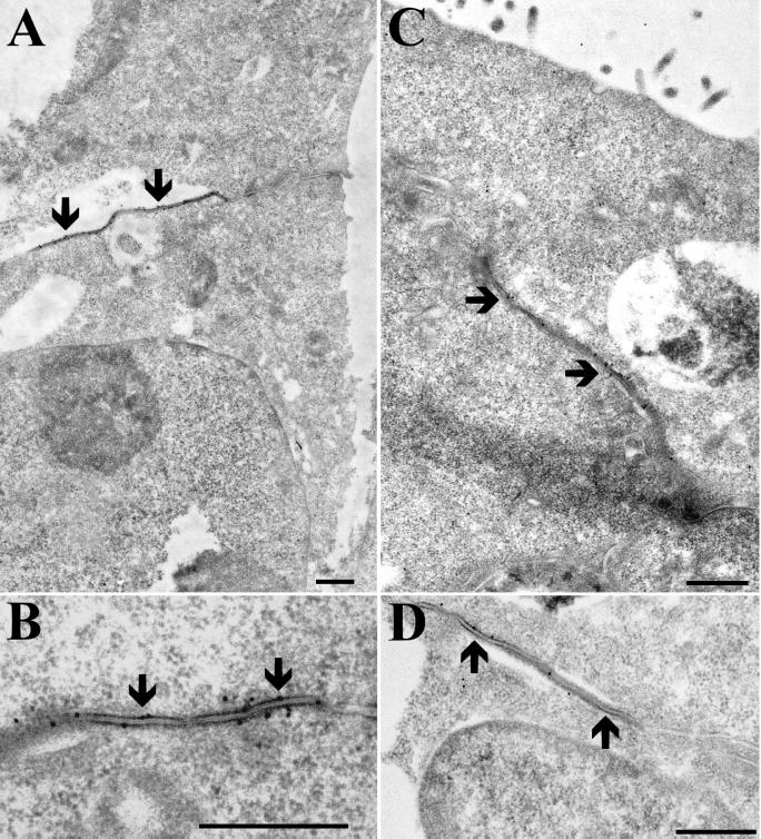
Figure 6.
Ultrastructural localization of GFP and Cx43 in MDCK cells stably expressing Cx43-GFP. Thin sections of Lowicryl-embedded, Cx43-GFP-expressing MDCK cells were immunogold labeled for GFP (A and B) or Cx43 (C and D). Note the specific localization of gold particles to the gap junction plaques (arrows). Bar, 0.5 μm.

