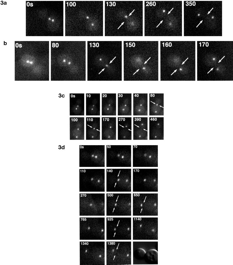
Figure 3.
In vivo analysis of spindle movements in wild-type and nip100Δ cells. Wild-type JKY85 (A and B) and nip100Δ (JKY86) (C and D) cells expressing Nuf2-GFP were observed by time-lapse fluorescence microscopy. Live cells were observed at 10-s intervals for up to 25 min. Elapsed times are shown are in seconds. Arrows denote the position of the bud neck. (A and B) Rapid penetration of mitotic spindle through the bud neck in wild-type cells. Note that wild type cells typically translocate their mitotic spindles through the bud neck within 3 min of the advent of anaphase spindle elongation. (C) Mutant nip100Δ cells generally do not translocate their spindles through the bud neck. Note that the spindle elongates to ∼4 μm within the mother but does not translocate into the daughter. However at ∼270 s after anaphase B begins, continuing spindle elongation causes the daughter spindle pole to pass through the bud neck. (D) A mother–daughter pair of nip100Δ cells, which fails to partition the spindle into the bud for an extended period. Note that, like the cells in C, the spindle has elongated to the mother cell cortex. However, the spindle remains in the mother for >14 min. However, at 1340 s, the spindle has elongated through the bud neck.

