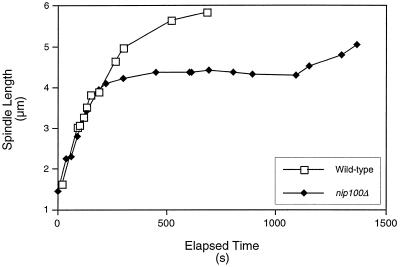
Figure 4.
Kinetic analysis of spindle elongation in nip100Δ mutants. Kinetic diagrams of wild-type (open squares) and nip100Δ (filled diamonds) cells. The nip100Δ cell pair is shown in Figure 3D. Fast elongation for both cells (0–200 s; spindle length, <4 μm) occurs at 0.89 μm/min. Slow elongation (200–1500 s) occurs at ∼0.20 μm/min in both wild-type and nip100Δ mutant cells. However, elongation pauses for >10 min (∼300–1100 s) in the nip100Δ cell while the spindle is stuck within the mother cell. Rates were determined as the slope of a linear curve fit of the data points in each phase.

