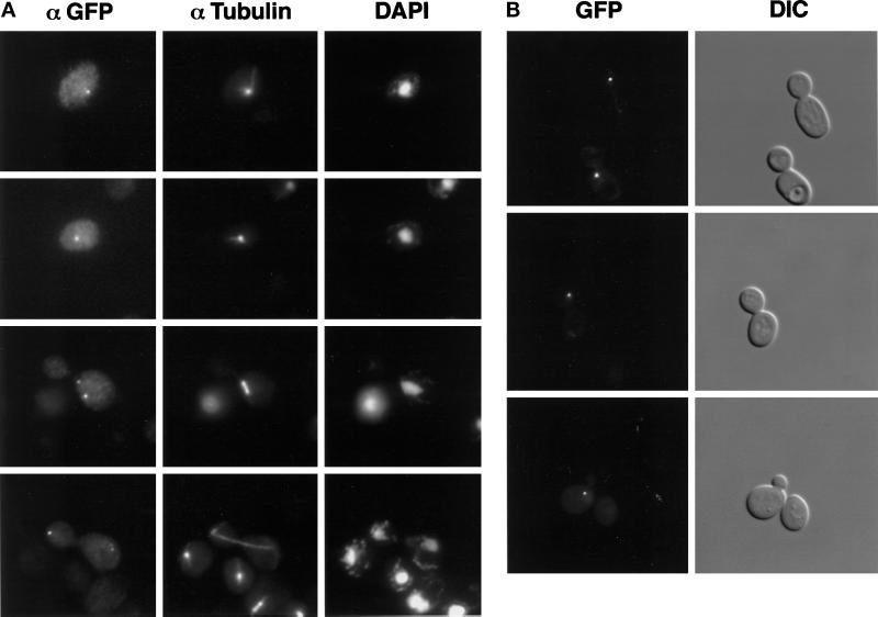
Figure 7.
Nip100 localizes to the spindle poles of vegetatively growing cells throughout the cell cycle. (A) Strain JKY77 (nip100Δ::HIS3) overexpressing Nip100-GFP from a 2μ plasmid was grown to midlogarithmic phase and processed for immunofluorescence. Nip100-GFP was visualized with a polyclonal antibody against GFP followed by FITC-conjugated secondary antibodies. Microtubules were visualized with a monoclonal antibody against α-tubulin followed by Texas Red-conjugated secondary antibodies. Chromatin was visualized with DAPI. (B) Pulse–chase expression of Nip100-GFP using the GAL1–10 promoter. Nip100-GFP was expressed for 30 min using 2% galactose and then repressed for 3 h using 2% glucose as the sole carbon source. In 58% of large-budded cells, Nip100-GFP is predominantly localized to the SPB, which is in the daughter cell (top two panels). In small and medium (preanaphase B) cells, Nip100-GFP is typically associated with a single SPB at the bud neck (bottom panel).

