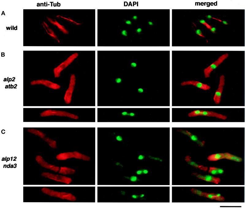
Figure 2.
Microtubule staining in ts alp2 (atb2) and alp12 (nda3) cells. Mutant and wild-type cells were prepared as in Figure 1, fixed in methanol, and stained with anti-tubulin antibody (TAT-1, left panel) or DAPI (middle panel). Merged figures are shown in the right panel. Representative figures are shown for wild-type (top row), ts alp2 (atb2) (second and third rows), and alp12 (nda3) (fourth and fifth rows).

