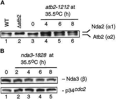
Figure 5.
The level of tubulin proteins in ts atb2 and nda3 mutants. (A) Total cell extracts were prepared from wild type (HM123, lane 1; Table 1), deleted atb2 (Δatb2, lane 2), or ts atb2-1212 (1212, lanes 3–6). Cells were cultured at 26°C in lanes 1 and 2. atb2-1212 cells were first grown at 26°C (lane 3) and shifted to 35.5°C. Samples were collected at 4 (lane 4), 6 (lane 5), and 8 (lane 6) h. Protein (2 μg) from cell extracts was run in SDS-PAGE, and immunoblotting was performed with anti-α-tubulin antibody (TAT-1). (B) ts nda3-1828 cells (DH12) were grown as described in A, and samples were taken every 2 h after the shift. Cell extracts (20 μg) were run in SDS-PAGE, and immunoblotting was performed with mouse anti-β-tubulin antibody (Sigma) (top) or anti-Cdc2 antibody as a loading control (bottom).

