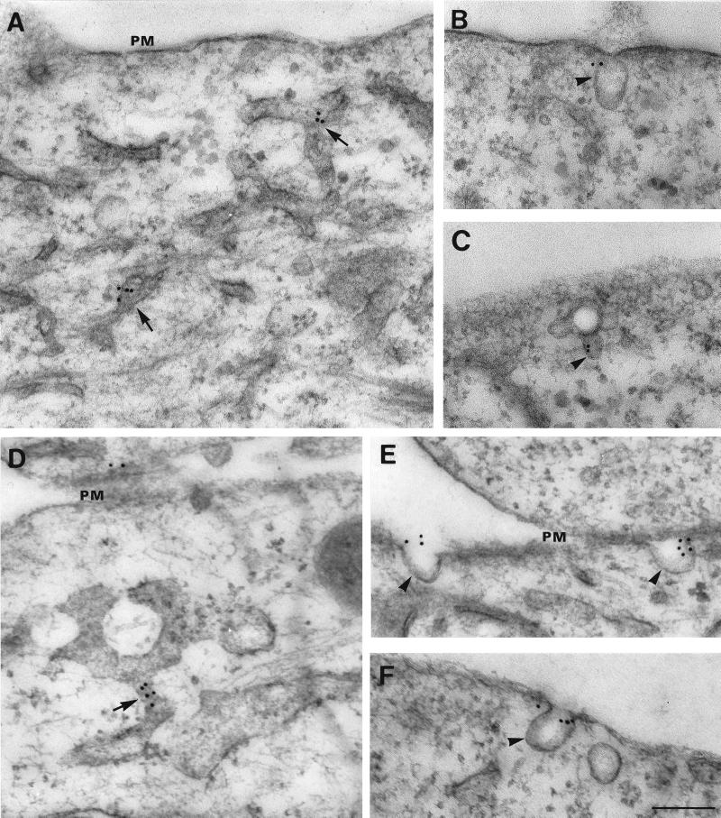
Figure 1.
Electron microscopic localization of AMF-R in NIH-3T3 fibroblasts and HeLa cells. HeLa (A, B, and C) and NIH-3T3 (D, E, and F) cells were postembedding immunolabeled with anti-AMF-R and 12-nm gold–conjugated anti-rat IgM secondary antibodies. Typical AMF-R labeling of smooth tubules (A and D, arrows) and cell surface caveolae (B, C, E, F, arrowheads) is shown. PM, Plasma membrane. Bar, 0.2 μm.

