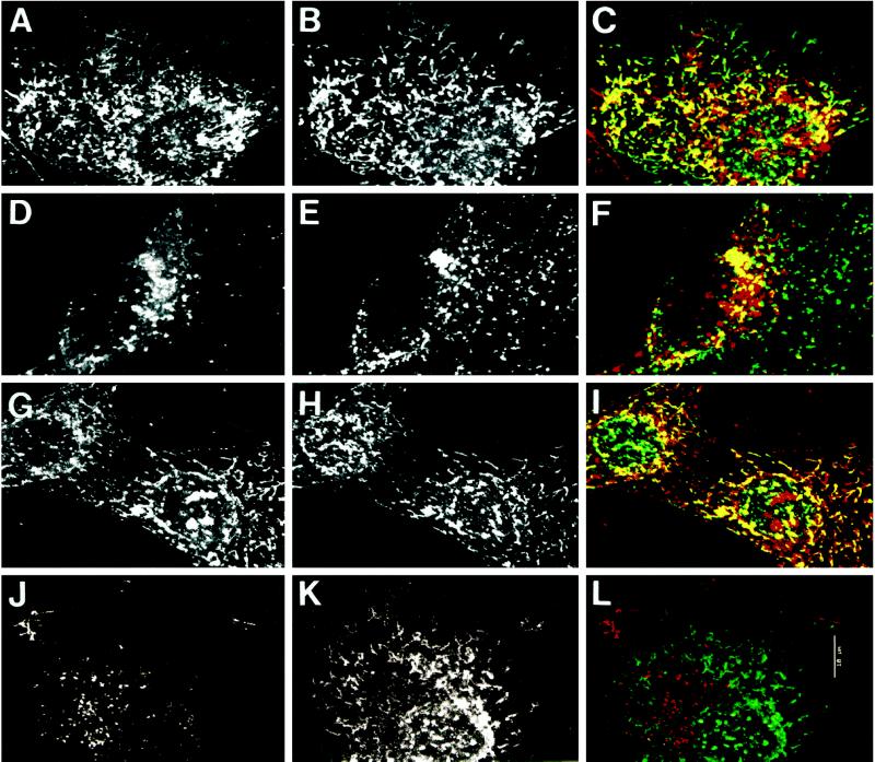
Figure 5.
Localization of internalized bAMF to AMF-R tubules by confocal microscopy. NIH-3T3 cells were pulse labeled with bAMF at 37°C for 1 h in regular medium (A–F) for 1 h in medium acidified to pH 5.5 to disrupt clathrin-mediated endocytosis (G–I), or in regular medium in the presence of 10-fold excess unlabeled AMF (J–L) before fixation with methanol/acetone. bAMF was revealed with Texas Red-streptavidin (A, D, G, and J) and AMF-R (B, H, and K) or LAMP-1 (E) labeled with the appropriate primary antibodies and FITC-conjugated secondary antibodies. Confocal images from both fluorescent channels were superimposed (panels C, I, and L, bAMF in red and AMF-R in green; panel F, bAMF in red and LAMP-1 in green) and colocalization appears in yellow. Bar, 10 μm.

