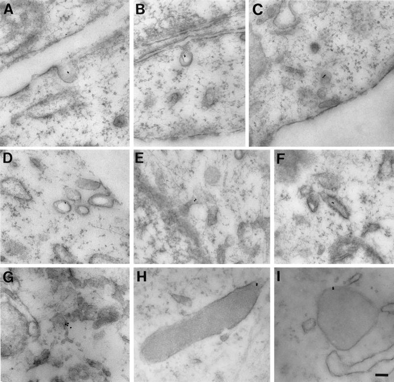
Figure 6.
Electron microscopy of the internalization pathway of bAMF. NIH-3T3 cells were pulsed with bAMF at 37°C for 10 (A, B, and H) or 30 min (C, D, E, F, G, and I). The localization of bAMF was revealed by postembedding labeling with 10-nm gold-conjugated streptavidin. After 10 min, bAMF is localized to cell surface caveolae (A and B). After a 30-min pulse, bAMF is localized to caveolae and smooth vesicles (C and D) and also appears in intracellular membranous tubules (E, F, and G) including distinctive smooth (E) and rough (F) ER elements. bAMF labeling of dense lysosomal structures is also detected (H and I). Bar, 0.1 μm.

