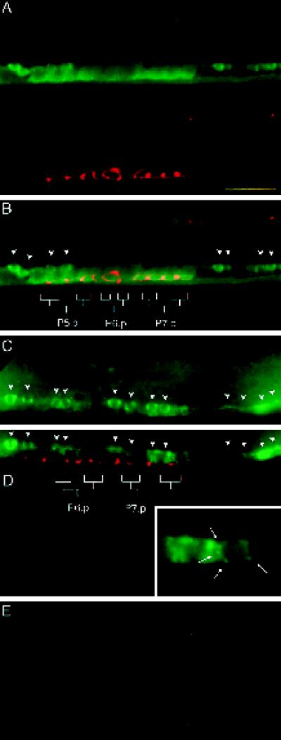
Figure 6.
LIN-10 is expressed in the developing vulva. (A–D) Wild-type, late L3 hermaphrodites. (A) Lateral view of the developing vulva. The upper panel shows staining with anti-LIN-10 antibodies (green), and the lower panel shows staining with the celljunction mAb marker MH27 (red). (B) Panels in A are merged. Positions of vulval epithelia (granddaughter cells of P5.p, P6.p, and P7.p) are indicated. Arrowheads indicate cell bodies of ventral cord neurons. Vulval epithelial cells can be distinguished from ventral cord neurons by their larger size and the presence of stained cell junctions (red). LIN-10 is present in the cytoplasm and at the plasma membrane. (C and D) Two focal planes showing the developing vulva and ventral cord neurons. Positions of ventral cord neurons are indicated by arrows. Some staining in D is from ventral cord neurons out of the plane of focus. Punctate, perinuclear LIN-10 staining can be seen in some vulval epithelial cells (D, inset, arrows). (E) lin-10(n299) late L3 hermaphrodite stained with anti-LIN-10 antibodies (green) as above. The region of developing vulva and ventral cord neurons is shown as in A–D. Bar, 10 μm.

