Figure 7.
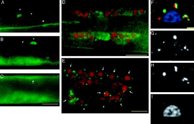
LIN-10 is expressed in neurons. (A–C) Wild-type, late L3 hermaphrodite stained with anti-LIN-10 antibodies (green). LIN-10 is present in ventral cord processes (A, *), lateral neural cell bodies and processes (A and B, arrowheads), and dorsal cord processes (C, arrow). Ventral staining in B and C is from the developing vulva out of the plane of focus. (D and E) Wild-type worms were stained with anti-LIN-10 antibodies (green) and DAPI (pseudocolored red), and images were taken with a DeltaVision wide-field microscope and deconvolved (see MATERIALS AND METHODS). (D) Lateral view of the nerve ring of an L4 hermaphrodite showing LIN-10 staining in nerve ring cell bodies and nerve ring neuropil. (E) Lateral view of an adult hermaphrodite tail showing punctate, perinuclear LIN-10 staining around neural nuclei (arrows). Some LIN-10 staining may also be associated with nonneural nuclei (arrowheads). (F) Merged image showing a neural cell body expressing transgenic ST-GFP and stained with anti-LIN-10 antibodies (red), anti-GFP antibodies (green), and DAPI (blue). (G) LIN-10 staining; (H) ST-GFP staining; (I) DAPI staining. Bars, 10 μm (A–E); 1 μm (F–I).

