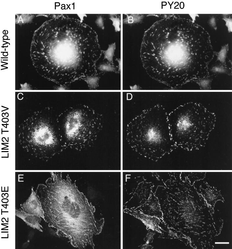
Figure 5.
Immunofluorescence analysis of the capacity and efficiency of paxillin LIM2 phosphorylation mutants to localize to FAs. CHO.K1 cells were transfected with avian paxillin cDNA containing mutations of LIM2. After 24 h of growth on glass coverslips in Ham’s F-12 media containing 10% FBS, ectopically expressed avian paxillin was visualized by immunofluorescence double-labeling with a chicken-specific, polyclonal antiserum Pax1 (A, C, and E) and a monoclonal antibody to phosphotyrosine, PY20 (B, D, and F). (A and B) Wild-type; (C and D) LIM2T403V; (E and F) LIM2T403E are representative of the transfected populations. Bar, 5 μm.

