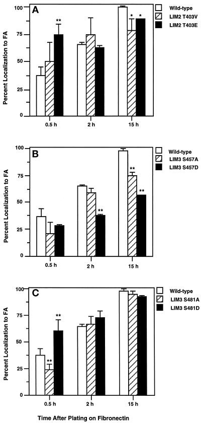
Figure 8.
Increased FA localization rate of paxillin molecules containing phospho-mimetic mutations of phosphorylatable residues within LIM2 or LIM3. (A) LIM2T403V and LIM2T403E. (B) LIM3S457A and LIM3S457D. (C) LIM3S481A and LIM3S481D. After adhesion to 10 μg/ml fibronectin-coated glass coverslips for 0.5, 2, or 15 h, the cells were processed and immunofluorescence double-labeled with Pax1 and PY20 or rhodamine-phalloidin. At each time point 150–200 transfected cells were counted with the number of avian paxillin transfectants displaying FA localization of the avian paxillin determined and divided by the total number of transfected cells. This is represented in bar graph form as the “Percentage localization to FA.” Four independent experiments were executed and tabulated to determine mean and SD from the mean. Statistical analyses were performed with Student’s t test; *, p < 0.05; **, p < 0.01.

