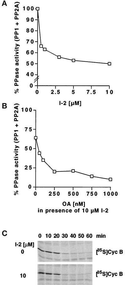
Figure 1.
The PP1 inhibitor I-2 does not prevent activation of mitotic cyclin B proteolysis. (A) Dose–response curve showing the effect of different concentrations of I-2 on the dephosphorylation of 32P-labeled glycogen phosphorylase A in Xenopus interphase extracts. (B) Dose–response curve showing the effect of different concentrations of OA on the phosphatase activity in Xenopus interphase extracts containing 10 μM I-2 (determined as in A). (C) The stability of 35S-labeled cyclin B was analyzed in extracts entering mitosis in the absence or presence of 10 μM I-2. Entry into mitosis was triggered by addition of nondegradable recombinant cyclin Δ90 at time zero, and samples taken at the indicated time points were analyzed by SDS-PAGE and phosphorimaging.

