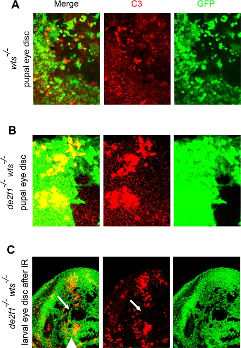
Figure 4. Loss of de2f1 does not affect resistance to DNA damage and pupal developmental apoptosis in wts mutants.
In all panels, clones were generated with the ey-FLP/FRT technique and mutant tissue is distinguished by the absence of GFP (green). (A–B) The pupal eye discs at 30 hr APF containing clones of wts mutant (A) and de2f1 wts double mutant (B) cells were stained with anti-Cleaved Caspase3 (C3) antibody (red) to detect apoptotic cells. In the pupal eye discs, developmentally regulated apoptosis is abundant in wild type cells (green) but is largely absent in wts mutant tissue (lack of green) and in de2f1 wts double mutant tissue indicating that de2f1 wts double mutant cells, like wts mutant cells, are protected from the cell death. (C) DNA damage induced apoptosis following irradiation was detected with anti-C3 antibody (red). There is an extensive apoptosis in wild type tissue. In contrast, de2f1 wts double mutant cells are protected from apoptosis after DNA damage. An example of de2f1 wts double mutant tissue is pointed by arrow. The morphogenetic furrow is marked by the arrowhead.

