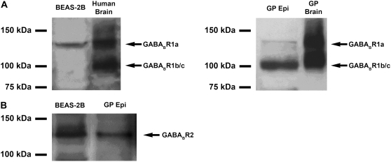
Figure 2.
Representative immunoblot analysis using protein prepared from (A, left) cultured human BEAS-2B cells (100 μg), whole human brain (25 μg), or (A, right) GP tracheal epithelium (Epi) (100 μg), and GP brain (25 μg). A larger splice variant (130 kD) corresponding to GABABR1a was detected in both brain-positive controls and in human BEAS-2B cells. A smaller splice variant (100 kD) that could represent either GABABR1b or 1c was detected in both brain-positive controls and in freshly isolated GP epithelium but not in cultured human BEAS-2B cells. (B) Representative immunoblot using antibody directed against GABABR2 identified an immunoreactive band of appropriate molecular mass (120 kD) in cultured human BEAS-2B cells and in freshly isolated GP epithelium. Images are representative of at least 3 independent immnoblot analyses from each cell or tissue source.

