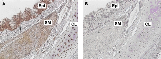
Figure 8.
Representative immunohistochemical staining of GAD65/67 in formalin-fixed GP trachea. (A) GAD65/67 immunostaining detected in tracheal epithelium (Epi). (B) Isotype-specific negative control primary antibody (rabbit IgG) in a parallel section of GP trachea. Both sections were counterstained with hematoxylin (original magnification: ×200). Images are representative of at least three independent immnohistochemical analyses from GP trachea.

