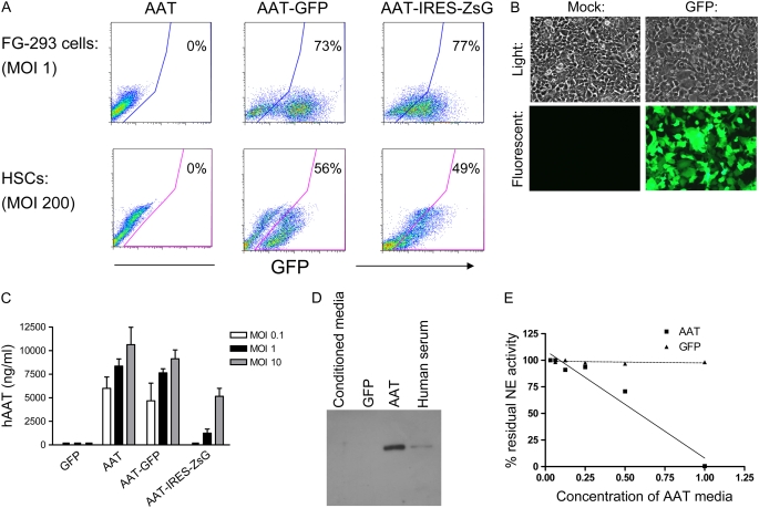
Figure 3.
In vitro testing of AAT-expressing lentiviral constructs. Cells infected overnight with each vector from Figure 2 were cultured further for 72 hours before analysis of gene expression. Separate aliquots were cultured in serum-free conditions for 48 hours before immunoblotting or anti-elastase assay. (A) Flow cytometry reveals approximately 75% of 293 cells and 50% of hematopoietic stem cells (HSCs) were transduced (GFP+ or ZsGreen+) with each vector. (B) Representative light and fluorescence microscopy shows GFP+ transduced 293 cells versus mock-infected controls. (C) Enzyme-linked immunosorbent assay (ELISA) of 293 cell supernatants reveals that cells transduced with “AAT,” “AAT-GFP,” or “AAT-IRES-ZsG” lentiviruses produce human AAT protein while cells transduced with virus encoding GFP alone do not. Increasing the MOI results in increased AAT protein levels. Error bars represent SEM. (D) Western blot of 293 cell supernatants after transduction with no virus (conditioned media), or lentivirus “AAT” versus “GFP.” hAAT secreted from transduced cells is the same size as hAAT in human serum. (E) Assay of anti-elastase activity in supernatants of 293 cells transduced with “AAT” vector versus “GFP” vector. “AAT” supernatant inactivated human neutrophil elastase (NE) in a dose-dependent fashion, while control “GFP” supernatant did not demonstrate anti-elastase activity.

