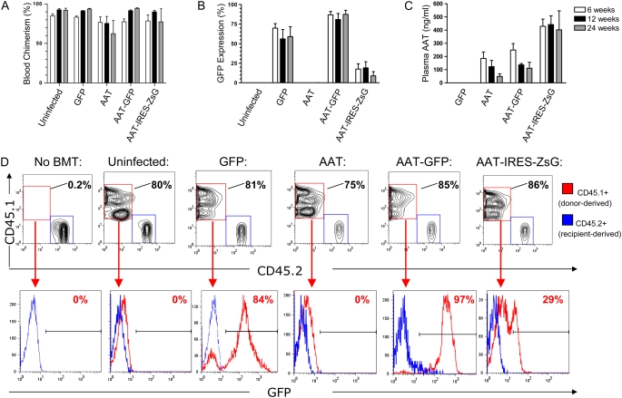
Figure 4.
Peripheral blood chimerism and in vivo gene expression after HSC infection and transplant. HSCs were infected overnight with each indicated lentiviral vector, and 3,000 HSCs were transplanted into each irradiated mouse recipient (n = 4 mice per group). Six, 12, and 24 weeks after transplantation, peripheral blood from each recipient was analyzed by flow cytometry to detect the percentage of donor-derived cells (blood chimerism; A) and the percentage of donor-derived cells expressing GFP or ZSGreen reporter genes (B). ELISA of blood plasma was used to quantify circulating human AAT protein levels (C). Error bars represent SEM. (D) Representative flow cytometry analysis from one mouse in each group, 24 weeks after HSC transplant: nucleated peripheral blood cells are analyzed after staining with antibodies that identify donor-derived cells (CD45.1) versus recipient-derived cells (CD45.2). A subgate of only donor-derived blood cells (CD45.1+/CD45.2−; represented as a red histogram) illustrates the percentage of donor-derived cells expressing the GFP or ZSGreen tracking reporter genes. The blue histogram overlay illustrates the absence of GFP expression in recipient cells (CD45.1−/CD45.2+) in each sample.

