Figure 5.
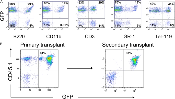
Assay of HSC functional capacity after lentiviral transduction. (A) Multilineage blood differentiation is illustrated by representative flow cytometry analysis of peripheral blood 14 weeks after transplantation of 3,000 HSCs infected with “AAT-GFP” lentivirus. Robust engraftment of differentiated lymphoid (CD3+ or B220+), myeloid (CD11b+ or GR-1+), and erythroid (Ter-119+) hematopoietic lineages derives from the transduced, transplanted donor cells (GFP+). Peripheral blood ELISA at this time point demonstrated an hAAT level of 149 ng/ml. (B) Analysis of peripheral blood drawn 13 weeks after primary transplant reveals 80% of all peripheral blood cells are donor-derived cells transduced with the “AAT-GFP” lentiviral construct (CD45.1+/GFP+; hAAT = 245 ng/ml). Twenty-four weeks after the initial transplant, 10 × 106 bone marrow cells from this primary recipient were transplanted into an irradiated secondary recipient. Seven weeks later, 83% of peripheral blood cells are derived from the original donor stem cells and continue to express both the GFP reporter gene (CD45.1+/GFP+).

