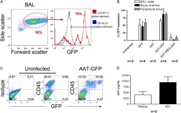
Figure 6.
Analysis of lung tissue 24 weeks after transplantation of HSCs infected with AAT-GFP lentivirus. (A) Flow cytometry analysis of cells obtained by BAL. Alveolar macrophages (AMs), representing 95% of BAL cells, are identified by their characteristic side scatter/forward scatter profile. Approximately 70% of AMs are GFP positive. (B) Percentage of cells expressing GFP/ZsGreen in the BAL, bone marrow, and peripheral blood compartments of recipient mice. No significant difference was found between tissue compartments in the percentage of cells expressing the tracking reporter gene (one way ANOVA > 0.05 for each vector). Error bars represent SEM. (C) Flow cytometry analysis of cells obtained by whole lung digestion. GFP+ cells are CD45+, indicating that transduced cells present in lung tissue are predominantly of hematopoietic phenotype. For negative controls, samples were prepared from a mouse transplanted with unmanipulated (uninfected) HSCs. Specificity of anti-CD45 staining is indicated by a sample exposed to nonspecific control antibody of identical isotype. (D) AAT expression in the plasma and epithelial lining fluid of recipients transplanted with HSCs infected with AAT-IRES-ZsG lentivirus.

