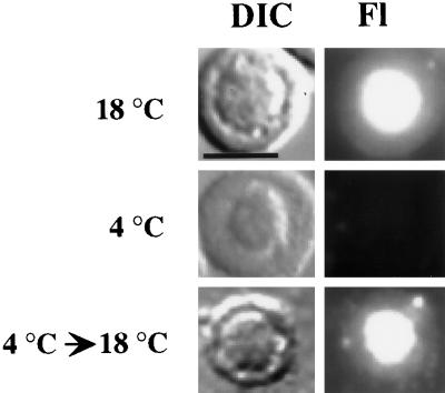
Figure 3.
Effect of temperature reduction on the nucleolar localization of U3 snoRNA. Fluorescently labeled, in vitro–synthesized U3 RNA was injected into nuclei of oocytes that were maintained either at 18°C (top) or at 4°C (middle) for 1 h before nuclear spread preparations were analyzed. In the temperature-shift experiment (bottom), the oocytes were shifted to 18°C for 1 h after being maintained at 4°C for 1 h. Each DIC image shows a single nucleolus, and the RNA signal is shown in the corresponding Fl image. Bar, 10 μm.

