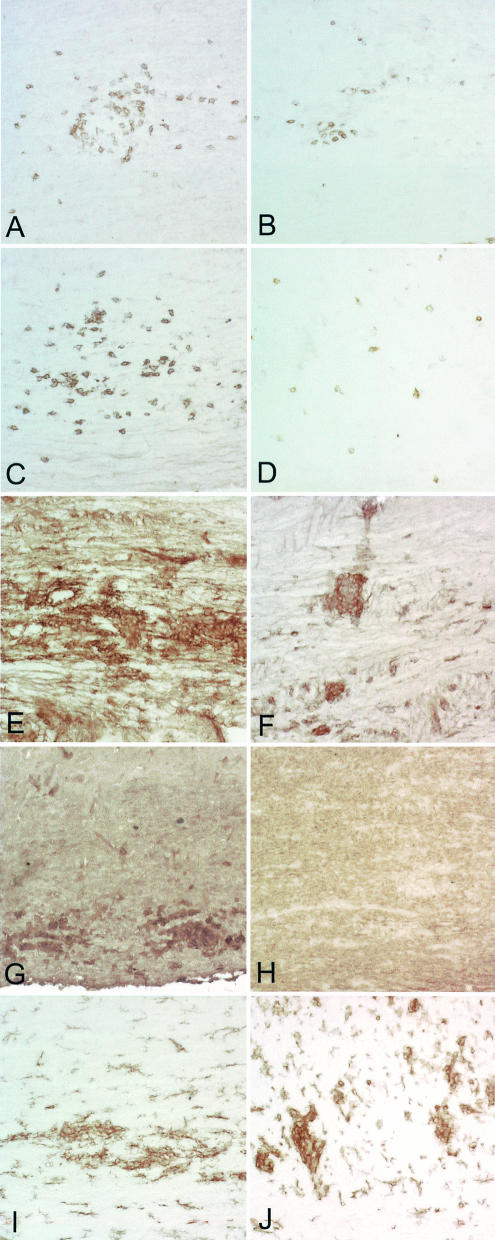
FIG. 4.
Decreased CD4, CD8, class I MHC, and class II MHC expression in spinal cords of IFN−/− H-2q mice. Immunoperoxidase staining for CD4+ T cells, CD8+ T cells, class I MHC, class II MHC, and F4/80 (macrophages) in spinal cords of IFN−/− H-2q and IFN+/+ H-2q mice was carried out at 12 days after infection. (A) CD4+ T cells are abundant in the spinal cord of an IFN-γ+/+ H-2q mouse. Some of the CD4+ T cells are present in the meninges, whereas others are present in the parenchyma, frequently in a perivascular location. (B) There are fewer CD4+ T cells in the spinal cord of an IFN-γ−/− H-2q mouse than in comparable areas of the spinal cord of an IFN-γ+/+ mouse (panel A). (C) A serial section from an IFN-γ+/+ H-2q mouse (panel A) was stained for CD8+ T cells. Note that the cells are distributed widely in the spinal cord parenchyma and have a distinct distribution compared to CD4+ T cells. (D) A serial section from an IFN-γ−/− H-2q mouse (panel B) shows that there are fewer CD8+ T cells than in an IFN-γ+/+ H-2q mouse (panel C). (E) Class I MHC-positive cells are present almost exclusively in the gray matter of the spinal cord of an IFN-γ+/+ H-2q mouse. (F) There is less expression of class I MHC in cells of the gray matter of an IFN-γ−/− than in an IFN-γ+/+ H-2q mouse (panel E). (G) A serial section (obtained from the sample shown in panel E) shows the localization of class II MHC-positive cells in an IFN-γ+/+ H-2q mouse. The cells are widely scattered in the gray matter and in the white matter. (H) A serial section (obtained from the sample shown in panel F) shows less expression of class II MHC-positive cells in the spinal cord of an IFN-γ−/− mouse than in that of an IFN-γ+/+ mouse. (I) Staining in an IFN-γ+/+ H-2q mouse for F4/80 demonstrates labeling of macrophages and a subpopulation of microglia. (J) An extent of F4/80 staining similar to that shown in panel I was observed in an IFN-γ+/− mouse.

