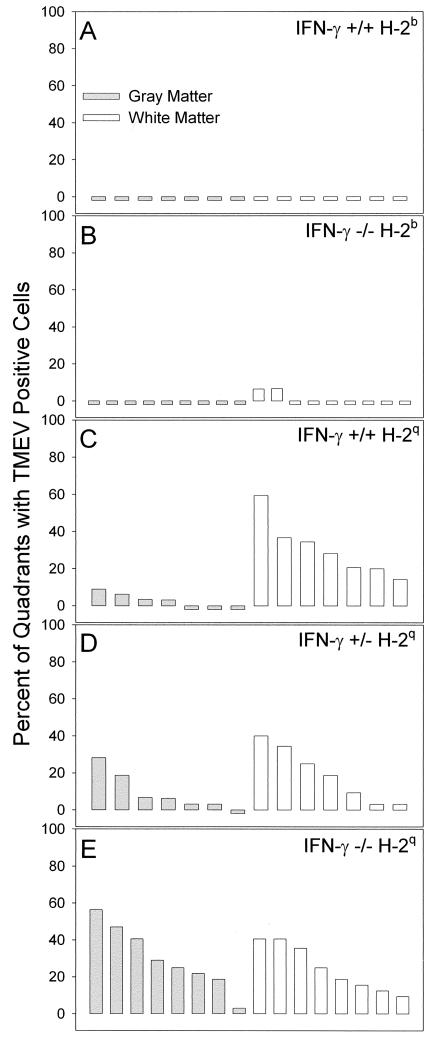
FIG. 7.
Quantitation of spinal cord quadrants with TMEV-infected cells in the gray matter and white matter of the spinal cord. The number of virus antigen-positive cells was determined by immunoperoxidase staining and expressed as the percentage of spinal quadrants showing virus antigen-positive cells in either the gray matter or the white matter. Analysis was done 16 days after infection. Each bar represents one animal. (A) No virus-antigen positive cells were observed in the gray matter or the white matter of normally resistant IFN-γ+/+ H-2b mice. (B) In contrast, there were small numbers of virus antigen-positive cells in the white matter of two of nine IFN-γ−/− H-2b mice but no virus antigen-positive cells in the gray matter. (C) A few virus antigen-positive cells were observed in four of seven IFN-γ+/+ H-2q mice, whereas multiple spinal cord quadrants showed virus antigen-positive cells in all seven of seven IFN-γ+/+ H-2q mice. (D) Similar distributions of virus antigen-positive cells were observed in IFN-γ+/−H-2q mice and IFN-γ+/+ H-2q mice. (E) In contrast, there were more spinal cord quadrants with virus antigen-positive cells in the gray matter of IFN-γ−/− H-2q mice than in those of IFN-γ+/+ H-2q mice (P = 0.001, as determined by the Student t test) or in those of IFN-γ+/− H-2q mice (P = 0.015, as determined by the Student t test).

