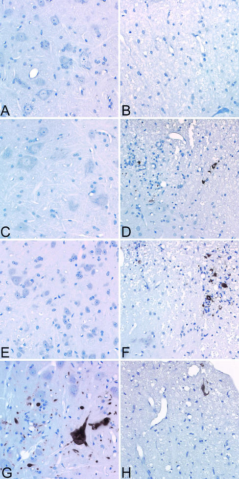
FIG. 8.
TMEV infection of gray matter and white matter cells in the spinal cord. Virus antigen staining was performed 16 days after TMEV infection. (A) Absence of virus antigen in anterior horn cells of the spinal cord of an IFN-γ+/+ H-2b mouse (B) Absence of virus antigen in the white matter of an IFN-γ+/+ H-2b mouse. (C) Absence of virus antigen in anterior horn cells of the spinal cord of an IFN-γ−/− H-2b mouse. (D)Virus antigen in the white matter of an IFN-γ−/− H-2b mouse. (E) Absence of virus antigen in anterior horn cells of an IFN-γ+/+ H-2q mouse. (F) Presence of virus antigen in the white matter of an IFN-γ+/+ H-2q mouse. (G) Presence of virus antigen in the gray matter of an IFN-γ−/− H-2q mouse. Virus antigen was localized predominantly in the cytoplasm of neurons. No similar virus antigen-positive anterior horn cell neurons were identified in infected IFN-γ+/+ H-2q or IFN-γ+/− H-2q mice. (H) Virus antigen localized to the spinal cord white matter of an infected IFN-γ−/− H-2q mouse.

