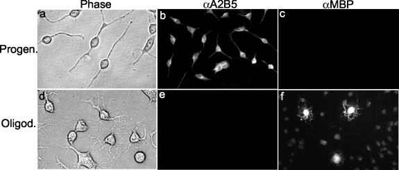
FIG. 1.
Morphological and biochemical properties of the undifferentiated CG-4 progenitor cells and the differentiated oligodendrocytes. (a to c) Progenitor cells were cultured in the undifferentiating medium as described in Materials and Methods. (d to f) Mature oligodendrocytes were differentiated from progenitor cells for more than 48 h in the differentiating medium (see Materials and Methods). Panels a and d are phase-contrast microscopic images of the cell cultures. Panels b and e and c and f show immunofluorescence staining results with the primary monoclonal antibodies to A2B5 (a marker for progenitor cells) and MBP (a marker for mature oligodendrocytes), respectively, and the secondary antibodies conjugated with FITC. Cells were observed under the microscope (Olympus IX-70), and all images were taken with the attached digital camera (MagnaFire).

