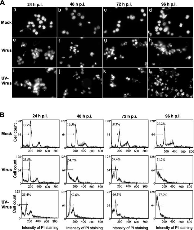
FIG. 3.
MHV-induced apoptosis of mature rat oligodendrocytes. Mature oligodendrocytes were mock infected (Mock) or infected with MHV-JHM at an MOI of 10 (Virus) or with UV-inactivated MHV-JHM at an equivalent MOI (UV-Virus). At the indicated time points (hours) p.i., total cells were collected and stained with propidium iodide. A portion of the stained cells was subjected to microscopic examination (A), and the remainder was used for flow cytometric analysis (B). (A) Morphological characteristics of the nuclei of oligodendrocytes. All photographs were taken with a digital camera (MagnaFire) attached to a fluorescence microscope (Olympus IX-70) with the same magnification. (B) Results of flow cytometric analysis. The bar in each graph represents the sub-G0/G1 population of cells (indicated as a percentage) that have the lowest intensity of propidium iodide staining. Data are representative of at least three independent experiments.

