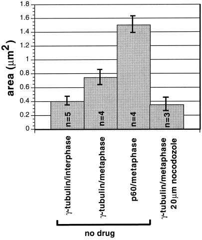
Figure 11.
Quantitative analysis of the cell cycle dependence and microtubule dependence of the centrosomal localization of katanin and γ-tubulin in Xenopus A6 cells. The area of concentrated p60 katanin staining and γ-tubulin staining at interphase centrosomes and metaphase spindle poles was measured (see MATERIALS AND METHODS) in several cells either untreated or treated with 20 μM nocodazole for 60 min to depolymerize microtubules. Averages of measurements in each category are shown. p60 katanin is not found at interphase centrosomes or nocodazole-treated spindle poles in these cells.

