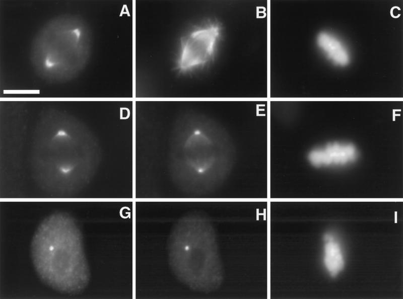
Figure 8.
Immunolocalization of p60 and p80 katanin at metaphase spindle poles in human fibroblasts. MSU1.1 cells were fixed and stained with a human p80 antibody (A), a human p60 antibody (D and G), a β-tubulin antibody (B), a γ-tubulin antibody (E and H), and DAPI (C, F, and I). p80 is shown concentrated at spindle poles relative to microtubules (A, B, and C), and p60 is shown colocalizing with γ-tubulin at spindle poles (D, E, and F). The concentration of p60 and γ-tubulin at spindle poles remains after microtubules are depolymerized with 20 μM nocodazole (G, H, and I). The area occupied by p60 in the presence of microtubules is greater than that occupied by γ-tubulin (D and E), whereas p60 and γ-tubulin occupy the same area after microtubules are depolymerized (G and H). Bar, 5 μm.

