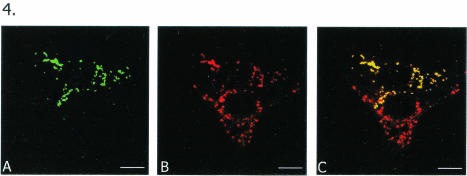
FIG. 4.
Morphologic similarities and cytoplasmic localization of VLS formed by GFP-NSP5 and infection-associated viroplasms in two adjacent cells at the light microscopic level. MA104 cells were first infected with rotavirus SA11 and then transfected with pGFP-NSP5. (A) Top cell expresses green GFP-NSP5. (B) Bottom cell demonstrates infection-associated viroplasms immunostained in red by anti-NSP5 antibody. Anti-NSP5 antibody also stained GFP-NSP5 in red in the top cell; this cell may or may not have been infected with the virus—most likely not, given the low MOI (1) coupled with a low efficiency of transfection, making a cotransfected-infected cell a rare occurrence. (C) Overlay shows GFP-NSP5-associated VLS in yellow due to the dual staining (green and red) of the chimeric protein in the top cell, whereas the infection-associated viroplasms remain red. Note morphologic similarities between VLS and viroplasms as well as their similar locations in adjacent cells. Bars, 10 μm.

