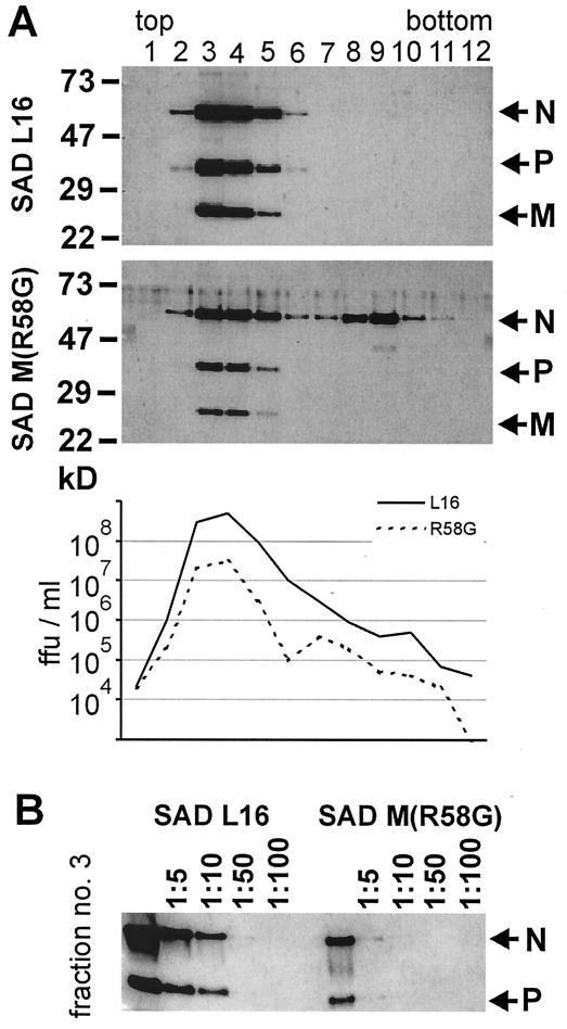
FIG. 5.
Protein content and infectivity of SAD M(R58G) virions. Two days after BSR cells were infected at an MOI of 1, cell culture supernatants were prepared and purified by centrifugation through 20% sucrose. After 18 h of centrifugation in a 10 to 40% iodixanol density gradient, 12 fractions (1 ml each) were collected. (A) The infectious virus titers were determined by end point dilutions, and the protein content of each fraction was analyzed in Western blots with sera recognizing RV N, P, and M proteins. ffu, focus-forming units. (B) To compare the amount of virus protein, virus peak fraction 3 was diluted, and N and P proteins were detected by Western blotting. Nearly 10-fold-less protein was detected in the supernatant from SAD M(R58G)-infected cells.

