FIG. 5.
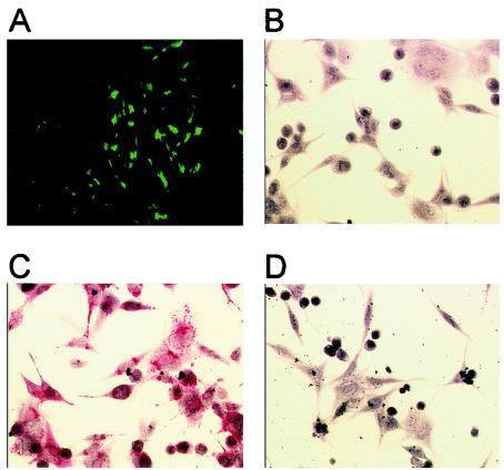
Fluorescence and immunohistochemistry analysis of wt-PCMV and recombinant PCMV infection of PMEC. (A) Peromyscus cells 5 days after infection with recombinant PCMV(ΔP33:G1EGFP). Magnification, ×320. (B) PMEC infected with recombinant PCMV probed with nonspecific negative antibody control. (C) PMEC infected with recombinant PCMV probed with recombinant anti-SNV human antibody. (D) PMEC infected with wt-PCMV probed with recombinant anti-SNV human antibody. In panels B to D, primary antibody incubations were followed by incubation with alkaline phosphatase-conjugated secondary antibody-colorimetric development. Magnification, ×640.

