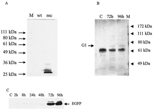
FIG. 6.
Western analysis of recombinant proteins expressed with recombinant PCMV virus. (A) Western blot of wt-PCMV-infected PMEC and PCMV(ΔP33:G1EGFP)-infected PMEC at 96 h postinfection with a monoclonal antibody to EGFP. The predominant band on the blot is EGFP protein with a molecular mass of ca. 27 kDa. (B) Western blot of SNV expression in PCMV(ΔP33:G1EGFP)-infected PMEC at 72 and 96 h postinfection with a rabbit anti-peptide antibody raised against an epitope of SNV-G1. The minor band at ca. 72 kDa corresponds to SNV-G1. (C) Western blot of time course expression of EGFP in PCMV(ΔP33:G1EGFP)-infected PMEC.

