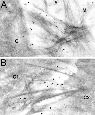
Figure 2.
Electron microscopy of CMP filamentous networks. Chondrocytes were cultured in the absence of ascorbate to prevent collagen secretion, before immunogold electron microscopy (see Figure 5I for the absence of extracellular Col II in the culture without ascorbate). (A) CMP filaments connect a chondrocyte to matrix. Notice the gold particles decorate the matrix filaments protruding from the cell membrane. C, cell; M, matrix; V, matrix vesicles. Arrows point to some of the gold particles coupled to a mAb against CMP. Bar, 120 nm. (B) CMP filaments connect a chondrocyte to another chondrocyte. C1, cell 1; C2, cell 2. Notice the gold particles decorate the matrix filament bundles connecting neighboring cells. Bar, 120 nm. See Figure 3C for higher magnification of gold-labeled CMP filaments.

