Figure 3.
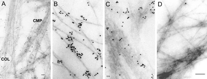
Immunogold electron microscopy. (A, B, and D) Chondrocytes were cultured in the presence of ascorbate to allow secretion of collagen fibrils. (C) Chondrocytes were cultured in the absence of ascorbate. (A) Two populations of filaments. COL, collagen fibrils; CMP, CMP filaments. Section reacted with a gold-coupled mAb against CMP. Notice the morphological difference between the two populations of filaments. Bar, 75 nm. (B) Collagen fibrils. Section reacted with a gold-coupled mAb against type II collagen. (C) CMP filaments. Section reacted with a gold-coupled mAb against CMP. (D) Control for immunogold electron microscopy. Section reacted with a gold-coupled mAb against type I collagen. Bar (B–D), 180 nm.

