Figure 1.
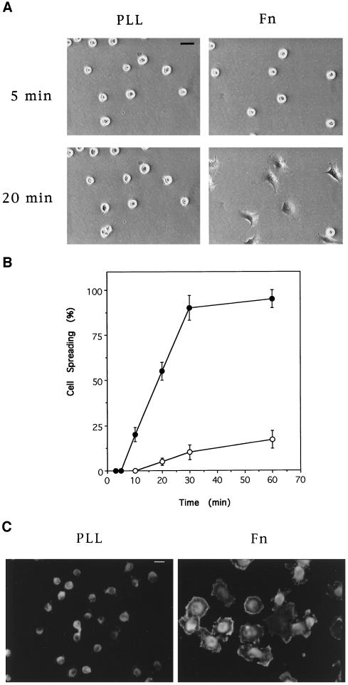
Cell spreading on Fn and poly-l-lysine. Quiescent cells in suspension were plated on coverslips coated with Fn or poly-l-lysine (PLL). Cells were fixed at the indicated times after plating and visualized by phase contrast microscopy (A) and scored for the percentage of spread cells (B) (•, Fn; ○, poly-l-lysine). (C) Twenty minutes after plating, cells were fixed and permeabilized, and filamentous actin was labeled with rhodamine–phalloidin; cells were then visualized by fluorescence microscopy. Bar, 20 μm.

