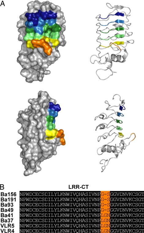
Fig. 2.
A model of the putative antigen binding surface of a BclA-CTD-specific VLR-B antibody. (A) Homology-based model of the tertiary structure of clone VLR4 constructed by using the crystal structure coordinates of hagfish VLR-B (PDB ID codes 2O6R and 2O6S) as a template. (Upper) Frontal view. (Lower) Side view. Hypervariable positions of each LRR module are indicated by color: LRR-NT (blue), LRR1 (light blue), LRRV (green), LRRVe (light green), CP (yellow), and LRR-CT (orange). (B) Alignment of the LRR-CT sequences of VLR-B clones specific for BclA-CTD. Invariant residues are shaded black, and variable residues are shaded orange.

