Fig. 3.
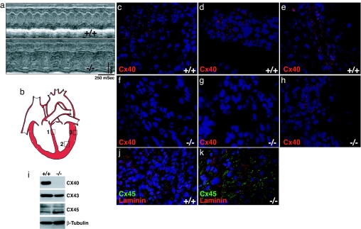
Impaired cardiac function in Dicer mutant hearts. (a) M-mode echocardiograms of P0 wild-type (+/+) and Dicer mutant (−/−) mice. (b) Diagram to show the areas of ventricles examined in confocal immunohistochemistry images: area 1 for c and f; area 2 for d and g; area 3 for e and h. (c–h) Confocal microscopic images for Cx40 protein expression (red) in the ventricles of P0 wild-type (+/+) and Dicer mutant (−/−) mice. DAPI stains nuclei. (i) Western blot analyses of connexin protein expression in P0 wild-type (+/+) and Dicer mutant (−/−) mice. β-Tubulin serves as a loading control. (j and k) Confocal microscopic images of Cx45 in P0 wild-type (+/+) and Dicer mutant (−/−) hearts. Laminin marks the cell surface and DAPI stains nuclei.

