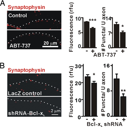
Fig. 3.
Bcl-xL is required for synapse formation. (A) Immunofluorescence microscopy for synaptophysin in axons of untransfected hippocampal neurons (DIV14) with or without 1 μM ABT-737 added at DIV5. Data were quantified as in Fig. 2C for three independent experiments (n = 6 cells per group). *, P < 0.05; ***, P < 0.001, Student's t test. White dots indicate the course of the axon. (B) Immunofluorescence microscopy for synaptophysin in axons of hippocampal neurons (DIV14) that were transfected (DIV5) with bcl-x hairpin RNA (pCAG-lacZ-shRNA-Bcl-xL) or control vector (pCAG-lacZ). Synaptophysin intensity (rfu) and number of puncta per 20 μm of axon in three independent experiments were quantified (mean ± SEM; n = 12 neurons per group). *, P < 0.05; **, P < 0.01; ***, P < 0.001, Student's t test.

