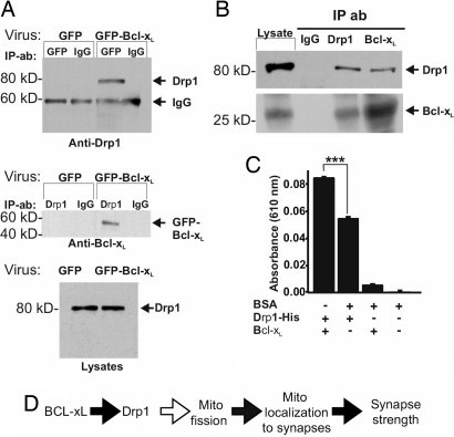
Fig. 6.
Bcl-xL and Drp1 interact. (A) (Top and Middle) Immunoblots of reciprocal coimmunoprecipitation experiments on hippocampal neurons (DIV13) transduced with viral vectors (DIV5) as indicated (n = 2). Antibodies used for precipitation (IP) and for immunoblots are indicated. (Top) Drp1 was coprecipitated with GFP-Bcl-xL, brought down by a GFP antibody. (Bottom) Blot of endogenous Drp1 in the starting lysates for immunoprecipitations above. (B) Immunoblots for endogenous Bcl-xL and endogenous Drp1 from adult rat brain after immunoprecipitation with the antibodies indicated. Each panel shows the starting lysate, control IgG precipitate, and the indicated precipitates probed with Drp1 and Bcl-xL antibodies. (C) Relative amounts of phosphate produced by GTPase activity in 1 h with purified recombinant proteins. Shown are results of three independent experiments (mean ± SEM). BSA had no GTPase activity above background, and this signal was subtracted from the other values. ***, P < 0.001. (D) Model of Bcl-xL-induced synaptic function. Black arrows indicate steps addressed in this work.

