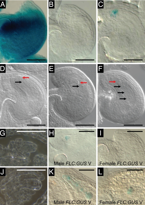
Fig. 2.
FLC:GUS is not expressed during female gametogenesis, and the paternally derived gene is expressed in the single-celled zygotes from vernalized plants. (A and D) Youngest open pollinated flower of nonvernalized Ler FRI + FLC-Col:GUS. (B and E) Youngest open pollinated flower of vernalized Ler FRI + FLC-Col:GUS. (C and F) Second youngest open pollinated flower of vernalized Ler FRI + FLC-Col:GUS. (A–C) GUS-stained viewed with DIC optics. (D–F) Cleared ovules for stage comparison. (G and J) Sections of C24 + FLC-C24:GUS through ovule primordia around the time of meiosis (G) and at the functional megaspore stage (J) viewed under dark-field conditions. (H and K) Ovules resulting from a cross between male Ler FRI + FLC-Col:GUS vernalized and female Ler FRI-Sf2 vernalized. (I and L) Ovules resulting from a cross between female Ler FRI + FLC-Col:GUS vernalized and male Ler FRI-Sf2 vernalized. (H and I) Single-celled zygote stage ovules (1 day after pollination). (K and L) Early-globular stage embryos (3 days after pollination). Black arrows, endosperm nuclei; red arrows, zygotic nuclei. (Scale bars: 50 μm.)

