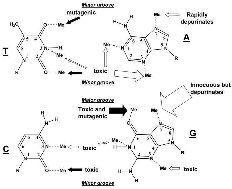
Figure 1.
Potential sites of chemical methylation in double strand DNA. The arrows point to each methyl adduct and whether the adduct is known to be predominantly toxic or mutagenic. The open arrows represent sites that are methylated by MMS, MNNG, and MNU. The filled arrows point to sites that are methylated by MNNG and MNU, but not detectably by MMS. Note that methylation of different sites on the same base at the same time is extremely rare. The size of the arrows roughly represent the relative proportion of adducts. In single strand DNA, the N1-adenine and N3-cytosine positions display a greater reactivity.

