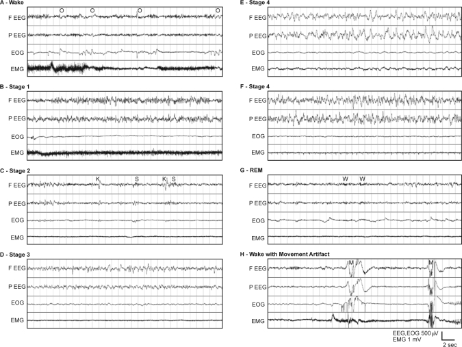
Figure 1.
(A) Example 30-sec epoch polygraph record showing stage Wake in a rhesus monkey. The appearance of high-frequency, low-amplitude EEG and high tonic EMG activity define stage Wake. EOG tends to show phasic activity representing rapid saccades and blinking. Ocular artifacts (O) are common in the frontal EEG. Slow movement artifacts often are shown on top of the high tonic activity in EMG. (B) Example polygraph showing stage 1 sleep in a rhesus monkey. (C) Example polygraph of stage 2 sleep in a rhesus monkey. K-complexes (K) and spindles (S) are shown in the EEG channels with slow waves occupying less than 20% of the 30-sec epoch. (D) Example polygraph showing stage 3 sleep in a rhesus monkey. High-amplitude slow waves occupy more than 20%, but less than 50%, of the 30-sec epoch. (E) Example polygraph of Stage 4 sleep in a rhesus monkey. High-amplitude slow waves occupy more than 50% of the 30-sec epoch. The slow waves resemble those of humans. The EEG slow waves caused some slow artifacts in the EMG of this example. (F) Example polygraph of stage 4 sleep in a rhesus monkey. High-amplitude slow waves occupy more than 50% of the 30-sec epoch. In some animals, higher frequency (>5 Hz) oscillations were commonly seen mixed with slow waves in both stage 3 and stage 4. (G) Example polygraph of REM sleep in a rhesus monkey. High-frequency, low-amplitude EEG appears accompanied by minimal EMG activity. Rapid eye movements are often seen as triangular waveforms in the EOG. Spindle-like wavelets (W) of about 20 Hz are common in EEGs of REM sleep. (H) Example polygraph showing movement artifacts in a rhesus monkey. These high-amplitude artifacts (M), appearing in all channels, are very common during active wakefulness.

